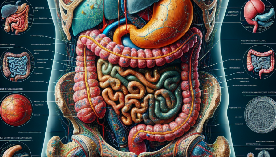
Welcome to our comprehensive guide on advanced abdominal imaging techniques. In this article, we will explore the various imaging modalities used to diagnose and evaluate abdominal conditions. From ultrasound to computed tomography (CT) and magnetic resonance imaging (MRI), we will delve into the advantages and limitations of each technique.
Ultrasound is often the first imaging modality used for abdominal evaluations due to its non-invasive nature and lack of ionizing radiation. It provides real-time images that can assess the liver, gallbladder, kidneys, and other abdominal organs. Additionally, ultrasound can be used to guide biopsies or drainages, making it a versatile tool in abdominal imaging.
CT scans, on the other hand, offer detailed cross-sectional images of the abdomen. By combining X-rays and computer technology, CT scans can provide information about the liver, pancreas, spleen, and gastrointestinal tract, among other structures. With the advent of multidetector CT scanners, faster acquisition times and improved image quality have revolutionized abdominal imaging.
MRI, with its superior soft tissue contrast resolution, is particularly useful for evaluating the liver, pancreas, and pelvic organs. It can provide valuable information about liver tumors, pancreatic masses, and pelvic pathologies. Furthermore, MRI does not involve ionizing radiation, making it a safe option for patients with repeated imaging needs.
Now, let's turn our attention to some of the advanced techniques used in abdominal imaging. One such technique is diffusion-weighted imaging (DWI), which measures the random motion of water molecules within tissues. DWI can help differentiate between benign and malignant lesions and detect early signs of tumor response to treatment.
Another advanced technique is magnetic resonance cholangiopancreatography (MRCP), which focuses on imaging the biliary and pancreatic ducts. MRCP can visualize gallstones, strictures, and other abnormalities within these ducts, aiding in the diagnosis and management of biliary and pancreatic disorders.
Lastly, we have contrast-enhanced imaging, which involves administering a contrast agent to highlight specific structures or abnormalities. Contrast-enhanced CT and MRI can provide valuable information about blood vessels, liver lesions, and other abnormalities that may not be clearly visible on non-contrast images.
In conclusion, advanced abdominal imaging techniques offer a comprehensive approach to diagnosing and evaluating abdominal conditions. From ultrasound to CT and MRI, each modality has its own strengths and limitations. By utilizing these techniques and their advanced variations, radiologists can provide accurate and detailed assessments of abdominal pathologies. Stay tuned for more in-depth articles on each imaging modality and its applications in abdominal imaging.…